Dr Mark Ross was among our first Ignition Fund awardees in 2024 and used the funding to undertake a secondment to the University of Birmingham to learn how to isolate immune cells from muscle tissue. He says: “This training opportunity was both an exciting and humbling experience – akin to being a PhD student again – learning new techniques from experts in the field.”
You can read Mark’s report here:
Isolating immune cells from muscle tissue to understand immune-muscle communication in ageing skeletal muscle
Dr Mark Ross, Institute of Life and Earth Science, Heriot-Watt University
Since completing a PhD in the early 2010s, my research has focused on how ageing affects circulating immune cells – particularly how exercise and physical activity can modulate ageing of the immune system. As we get older, we have more circulating pro-inflammatory immune cells, which are linked to the development and progression of cardiovascular disease, and may even be implicated in neurological conditions, such as dementia. As a result, understanding the role of the immune system in age-related diseases is essential to identify therapeutic targets for prevention and/or treatment.
We know from previous work that older adults exhibit greater number of immune cells in skeletal muscle than younger adults. This raised a compelling question – ‘do immune cells play a role in muscle mass loss and dysfunction with advancing age?’ which I am now focusing my research on.
Until now, my work has primarily examined immune cells in blood (namely circulating stem cells and angiogenic immune cells – which take part in vascular repair). To explore the role of immune cells in muscle ageing, I first needed to learn how to isolate them from muscle tissue. Thanks to the financial support from Vivensa and the expertise of my long-term collaborator Dr James Turner, who had performed this technique previously and published in this area, I visited the University of Birmingham for hands-on training. This experience would be vital for demonstrating technical competence in future research funding applications.
The Academy Ignition Fund covered my travel and accommodation expenses, bought laboratory reagents and consumables, enabling this exciting training opportunity. Without this support, gaining practical experience before submitting a major research funding application would have been difficult, and my lack of direct experience with the method could have raised concerns during peer review.
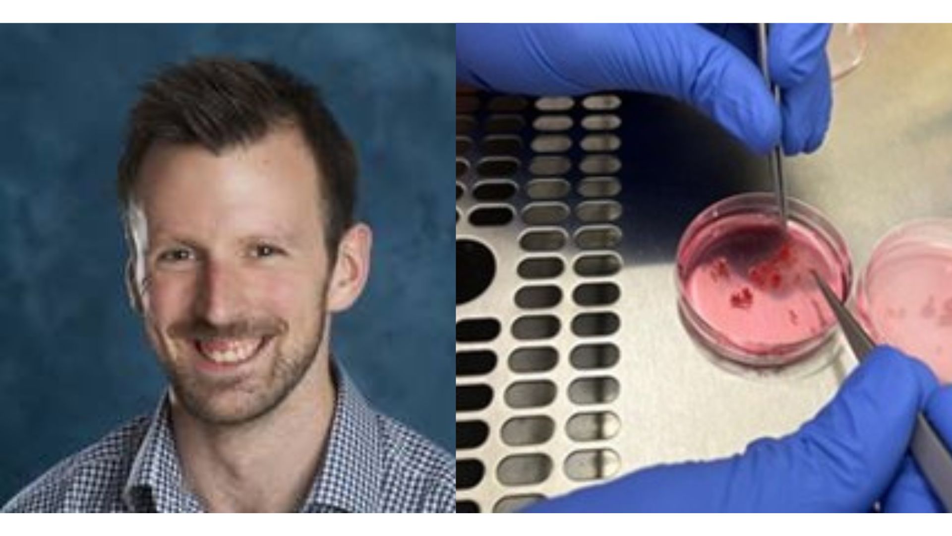
During the training, we collected muscle and then undertook an enzymatic digestion of the samples using different enzymes, allowing us to confirm the feasibility of the technique and also identify which enzyme yielded the best results. This experimentation added a layer of optimisation to the training which will strengthen our case for future research funding.
Personally, this training opportunity was both an exciting and humbling experience – akin to being a PhD student again – learning new techniques from experts in the field. The optimised method, and the data from this work, is supporting several research funding applications for submission later this year, which would not have been possible without the Vivensa Foundation’s support.

Figure. A human skeletal muscle sample (top left) which was then minced and digested (bottom left, top right) to then undertake flow cytometric analysis to quantify the immune cells, and markers of immune cell function (bottom right).
Acknowledgements: I would like to thank Professor Leigh Breen (University of Leicester), Dr Marie Korzepa and Mr Archie Belfield (University of Birmingham) for their support in collecting the muscle biopsies as part of this work.